Scientific and professional activity
Zacharias Chalampalakis studied Physics at the Kapodistrian University of Athens, Greece from where he graduated in 2010. After completing a Master’s degree in Medical Physics from the University of Aberdeen in 2012 he joined the UK national school of healthcare science as a trainee medical physicist. In 2015 he successfully completed the training scheme and became a registered clinical scientist (medical physics) specialising in imaging with ionising radiation. As part of his training he also successfully acquired a Master’s degree in Clinical Science from the University of Liverpool. From 2015 to 2018 he worked as a medical physicist at the PET Centre of King’s College London, supporting clinical scanning and assisting in research studies. There he gained an interest in PET, PET/MR and medical image reconstruction which driven him to pursue his PhD studies in the field of PET/MR reconstruction at the BioMaps laboratory.
His interests lie in the areas of medical image analysis, medical image reconstruction techniques and development of software for medical imaging.
PhD topic: Modelling and Reconstruction of a 3-D Whole Body parametric map in Hybrid PET-MRI Pharmacological imaging
His PhD topic is focused on whole-body parametric imaging using PET/MR datasets.
PET (standing for Positron Emission Tomography) is a highly sensitive method for molecular imaging, which makes use of specially radiolabelled tracers that target specific molecules and physiological functions in the body. Those act as probes that can be detected by the PET scanner and can be used to image and monitor the targeted molecule’s pathways in the body.
From the introduction of radioactive tracer in the body (almost always via an injection) to its clearance, the tracer distribution changes with time. Capturing this information using a PET scanner over a sufficient amount of time allows for full characterization of the kinetic processes and provides valuable information on the probed molecule’s processes. Models can be designed to describe the kinetic processes of molecules based on some generic assumptions. These assumptions form the basis of pharmacokinetic modelling. The fitted parameters of the models of interest (which apply for the molecule/tracer in study) can be used to quantitatively compare processes between different regions/organs or even identify regions that relate to specific molecular activity. The same parameters can be estimated for every single imaging unit of a PET scan, which is the voxel. A common voxel size in PET imaging is normally of a few mm in size.
When the kinetic parameters of interested are calculated for every single voxel, an image can be created that allows for comparison or even detection of regions of interest that correspond to a specific activity of the tracer in study. This is referred to as parametric imaging, and these images as parametric map images.
Dynamic PET data are required over the whole body in order to create whole body parametric maps. With the exception of some recent manufacturing achievements which allowed production of long axial field of view PET scanners, most scanners utilised clinically have an axial field of view between 15-25cm. This means that it is not possible to capture dynamic information form the whole body at once. To make use of these scanners for whole body dynamic imaging, dynamic protocols that repeat multiple acquisitions over the body at different axial positions (referred to as bed positions) are used. A diagram of a dynamic acquisition using 5 bed positions is shown below.
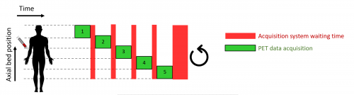
One of the disadvantages if this acquisition strategy is that the timing sampling is reduced resulting in reduced overall sensitivity and possible limitations in the modelling capabilities from the acquired datasets. The focus of this PhD research activities is to recover some of the lost sensitivity and improve parametric imaging for whole body protocols, using innovative reconstruction techniques.
Some of the latest findings of the PhD are described in a recent EANM presentation, which can be found here: https://www.youtube.com/watch?v=cQqs41GnwGI
And Poster!