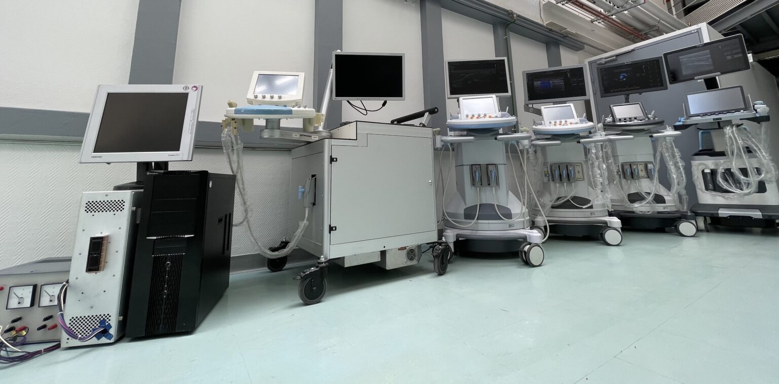
The core business of the group « methodological developments in ultrasound » is centered around ultrafast ultrasound imaging. Developed since the late 1990s, through extensive academic and industrial collaborations, it opens the doors of ultrasound to new quantification methods and new imaging biomarkers for medical diagnosis..
Ongoing projects:
Innovations in elastography to quantify viscosity and anisotropy of muscle: INNOVAN project
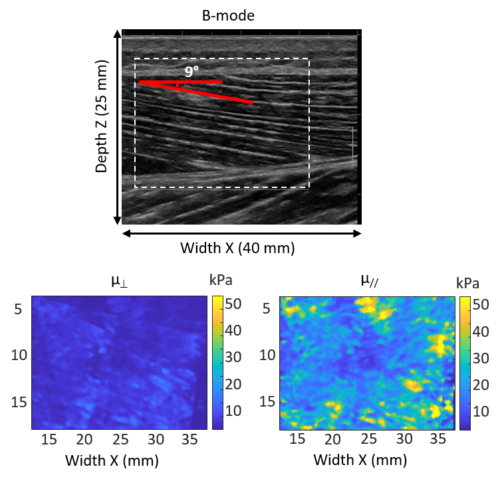
Ultrasonic (US) Shear Wave Elastography (SWE) has become popular in both basic sciences and clinical practice for measuring local mechanical properties of biological tissues in a non-invasive manner. However, for anisotropic and viscoelastic tissues such as muscles, SWE elastography suffers from two drawbacks which limit its application. 1) SWE 2D measurements are not able to take into account the muscular anisotropy (AN) due to fibers moving in 3D space, which induces a measurement bias. 2) Although viscosity is a crucial property for muscle, current SWE technologies do not allow viscosity to be measured. Therefore, the aim of the INNOVAN project is to tackle these two drawbacks, in order to greatly expand the potential of SWE. The ambition of this multidisciplinary project is to propose innovations in US imaging in order to 1) obtain a better fundamental understanding of muscle behavior and 2) to carry out important clinical applications. To this end, we have brought together a consortium of major research groups in physics, biomechanics, physiology and medicine.
Intramuscular FAT quantification by using UltraSound imaging: FATUS project
The first objective of the FATUS project is to lift the lock on the quantification of intramuscular fat content by ultrasound thanks to an innovative beamforming method. A validation step by MRI will confirm the achievement of this objective by developing specific sequences for fat quantification. By knowing and mapping these infiltrates, the third objective is to better understand the mechanical behavior of muscles in the presence of fat by improving ultrasound elastography techniques. The fourth objective is oriented around a characterization and a three-dimensional quantification of the muscles. The last objective is to provide a first clinical applications for the diagnosis of muscle sarcopenia due to aging. This multidisciplinary project therefore proposes to start from innovations in the field of ultrasonic medical imaging, to move towards a better fundamental understanding of the 3D behavior of the muscle, then towards clinical applications for a particularly important disease for old people. In order to carry it out, we have brought together a multidisciplinary team of researchers who are leaders in their fields (physics, industry, biomechanics, medicine).
Quantification of nonlinear elastic properties of tumors by US elastography and MRI: QUANTUM project
The objective of this study is to develop an experimental protocol of ERM and elastography-US which quantifies the mechanical properties of tumors grafted subcutaneously in small animals and subjected to uniaxial compression. We thus wish to have a better knowledge of the mechanical characteristics of tumors and to evaluate their link with the components of pressure (solid pressure, interstitial fluid pressure), but also their relationship with histological measurements (quantity of collagen, architecture).
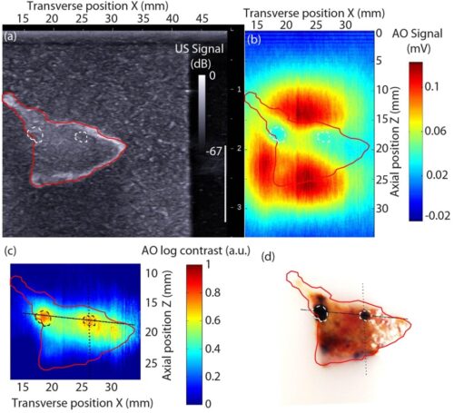
Acousto-optic imaging: AO project
Due to the multiple scattering of light within biological tissues, deep non-invasive optical medical imaging is very difficult. Acousto-optical imaging is a technique combining ultrasound and light which makes possible to recover optical contrast at depths of a few centimeters with millimeter resolution. Recent advances in acousto-optical imaging use short focused ultrasound pulses, often averaged over several hundreds or thousands of pulses. As the pulse rate of commercial probes is limited to about a few ultrasound cycles every 100 ms, acquiring an acousto-optical image typically takes several tens of seconds due to the high number of acoustic excitation pulses. We propose here new acousto-optical imaging techniques based on the use of ultrafast ultrasonic plane waves or ultrafast ultrasonic structured waves instead of ultrasonic focused waves which makes it possible to drastically increase the imaging rate.
J.-B. Laudereau et al., J. Biophoton, 2014
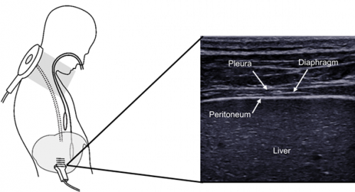
Preventing diaphragm dysfunction in the intensive care unit with innovative ultrasound biomarkers: ULTRADIAPH project
In the intensive care unit (ICU), mechanical ventilation relieves the work of respiratory muscles, especially the diaphragm, the main muscle for breathing. Mechanical ventilation ensures adequate gas exchange but can produce adverse effects that are associated with increased mortality, morbidity and health costs. The occupancy rate of intensive care beds in France is high (> 80%) and any strategy aimed at preventing an extended stay is crucial and more relevant than ever in the context of the COVID-19 pandemic. Avoiding insufficient or excessive diaphragm work has become an essential goal in the management of mechanically ventilated patients. This strategy, called protective diaphragm ventilation, cannot be implemented without reliable and easily accessible tools to assess the diaphragm, and more broadly, the accessory inspiratory and expiratory muscles. The monitoring of respiratory muscles in the ICU is complex and the reference methods based on the measurement of transdiaphragmatic pressure are invasive, not very available and require a great deal of expertise. In order to overcome these limitations, a new concept of ultrafast, ultraportable and dematerialized imaging technique has recently been developed. This approach induces a paradigm shift by going beyond the limits inherent in the miniaturization of hardware and opens up new horizons for image analysis, notably using artificial intelligence. This is a unique opportunity to implement high quality ultrasonic biomarkers in an ultraportable form factor. Building on this platform and on recent advances in ultrafast ultrasound, this project aims to develop an ultraportable solution specially designed for the assessment of respiratory muscle structure and function. We will use this new tool to improve knowledge of the structure and function of respiratory muscles, with the ultimate goal of optimizing the management of these ICU patients.
Past projects:
Towards and individualized periodization of training load adapted to muscle properties to reduce injury occurence in sprint running: FULGUR project – Website of the project
During the Olympics, running fast is the most common motor task, with the 100m sprint being one of the most anticipated events. Yet, achieving such running sprints requires both extreme athletic ability and a robust musculoskeletal system in order to limit the risk of injury. Muscle injuries of the lower limbs are indeed the main cause of interruption of training or competition on the international scene. In this context, France has the particularity of being historically strong in sports focused on speed and recognized for the quality of the research carried out for the understanding of sprint performance. This research program is carried out in close collaboration with the French athletics, rugby and ice sports (bobsleigh) federations. FULGUR brings together the world’s leading experts in muscle biomechanics, research on strength and conditioning, clinical imaging, health behaviors and machine learning applied to very high performance sports in order to pursue a triple objective:
• Evaluate the mechanics of the sprint at the level of the center of mass and the articulation segments in order to quantify the workload of the structures specific to these scales in real conditions; in this research new elastography parameters using ultrafast ultrasound imaging are being developed to prevent muscle injuries.
• Determine the individual musculoskeletal profile of the elite athlete in order to offer tailor-made strengthening programs to optimize the efficiency of the propulsion in the race;
• Estimate the level of risk of injury and propose individualized prevention content based on a multifactorial approach.
Video of the project report
Quantification of Mechanical, Anisotropic and Non-Linear properties of muscle in UltraSound Elastography: MALUS project
The objective of this project is to develop two innovative elastography methods in the transverse isotropic media to better characterize the muscles. 1/ a method for quantifying elastic non-linearity; 2/ a simple method of quantification of the anisotropy without movement of the probe for more ease of clinical use.
Minimalist and robust measurement of arterial diameter for a portable medical device: DIAMAND project
The measurement of physiological parameters is essential for the diagnosis of a chronic pathology or the monitoring of its evolution in response to a treatment. The evaluation of these parameters in daily life is a technological challenge for personalized medicine. In recent years, worn systems have multiplied, whether for medical purposes or for well-being. They perform well in terms of ergonomics and autonomy, but the disappointing robustness of the measurement remains problematic from a medical point of view. The approach taken by the DIAMAND project is to combine these different methods with an ultrasound measurement of physiological parameters, and in particular the diameter of the artery. The objective is to propose and test a minimalist architecture and acquisition strategy to obtain ultrasound artery diameter measurements with high precision (some μm). This « minimalist » aspect will make it possible to limit the overall size of the device, its energy consumption, and the quantity of data generated, in order to be able to subsequently integrate this measurement into a multimodal device for measuring physiological parameters.Minimalist and robust measurement of arterial diameter for a worn medical device: DIAMAND project
Multiparametric ultrasound imaging of diaphragm: RESPIMYO project
Led by Damien Bachasson from the Institute of Myology, this project in collaboration with BioMaps, aims to develop innovative and simple methods to characterize the diaphragm under complicated conditions in intensive care units.
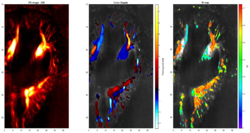
Kidney perfusion mapping by ultrafast ultraSound imaging for noninvasive diagnosis of chronic nephropathy: PERFUSION project
This project aims to define a new mapping of renal perfusion using ultrafast ultrasound imaging for the non-invasive diagnosis of chronic nephropathy. This research will combine two recent ultrasound techniques developed by us in recent years: elastography and ultrasensitive Doppler, in order to define a new vascularization outflow and resistivity mapping of transplanted kidneys.
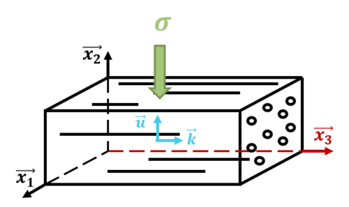
Elastic non-linearity in transverse isotropic medium: MUSCLOR project
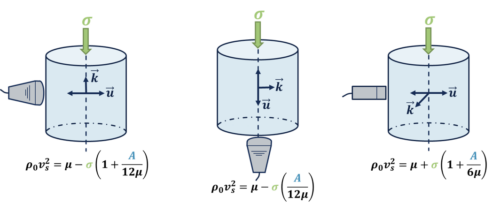
Recent developments in the field of elastography aim to quantify new mechanical properties of tissues, complementary to the shear modulus which describe the linear elastic properties of an almost incompressible medium. In this context, the measurement of elastic nonlinearity of tissues has recently been proposed on the basis of acoustoelasticity theory. Until now, most acoustoelastic theoretical developments have assumed that tissues are isotropic. However, this strong assumption does not hold in all human tissues, such as muscles which are generally considered to be transversely isotropic (TI). In this project, the theory of acoustoelasticity in an almost incompressible TI medium is developed and implemented experimentally on TI phantoms and on muscle tissues ex vivo and in vivo. In practice the evolutions of the speed of the shear waves is evaluated compared to the uniaxial static stress exerted on the medium giving access to the nonlinear elastic parameter A (some hundreds of kPa). This work opens the way to a better characterization of muscle mechanical properties as well as to the development of a new potential biomarker.
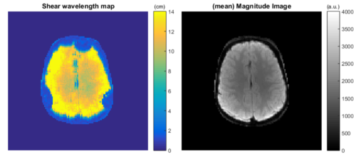
Passive elastography passive of the brain by using MRI: BPALP project
Elastography is a recent imaging technique (MRI or ultrasound (US)) which quantifies the biomechanical properties (BMP) of soft tissues. MR cerebral elastography has been widely studied recently and has made it possible, mainly ex vivo, to better understand tumor or degenerative pathologies. But this technique suffers from drawbacks for daily clinical practice. In ultrasound, due to the skull bone, it is not possible to non-invasively study brain BMPs without neurosurgery. In MRI, it is necessary to vibrate the head from an external actuator, which makes daily clinical diagnosis difficult. Thus, brain BMPs are poorly understood in vivo and are poorly used as a biomarker for cancer diagnosis or treatment monitoring against other organs. We propose to develop an original MRI method without an external vibrator for the diagnosis of brain cancer tumors with an innovative technique derived from ultrasound imaging, passive elastography. We develop MRI sequences: on clinical magnets, 3T for understanding BMPs and daily clinical practice; and preclinical, 7T for the fundamental understanding and follow-up of chemotherapy treatment. This is validated by passive ultrasound elastography, considered as gold standard, in preclinical with craniotomy and in humans during neurosurgery.