On this research theme, we are developing ultrasound monitoring methods for ultrasound-induced therapies. These combinations are based on the use of multimodal systems combining therapeutic ultrasound and another imaging method.
Ongoing projects:
Highly-SENSItive CAVitation Mapping for Advanced Monitoring of Ultrasound-Mediated Blood Brain Barrier Opening: SENSICAV project


Ultrasound localization miscroscopy of the Blood-Brain Barrier During Ultrasound Therapy of the Brain: SUPERBHE Project
For some years, a super-resolution ultrasound technique has been proposed in order to overcome the resolution limits imposed by conventional ultrasound. This approach, called ULM, proposes to exploit the reconstruction techniques used in optical imaging. Thus, the image obtained is no longer directly reconstructed from the signal backscattered by the microbubbles but various algorithms are implemented in order to follow the movement of these agents previously injected into the blood flow and to reconstruct a cartography of the vascular network with a resolution that can around 5 µm. Depending on the size of the vessels and the speed of the flow, tracking the movement of the microbubbles may require the acquisition of several tens of thousands of images over several seconds. The development of this approach has been possible thanks to plane wave imaging and the development of new ultra-fast ultrasound devices in recent years. We propose here to observe by super-resolution ultrasound the vascular variations induced by therapeutic ultrasound during the opening of the BBB.
Past projects:
Development and validation of a passive imaging device for acoustic cavitation through the human skull: CRANIUS Project
Treatment of numerous brain pathologies (glioblastoma, Alzheimer’s, Parkinson’s) remains a major challenge due to the presence of the blood-brain barrier (BBB) preventing access to the brain to most drugs. Ultrasound (US) associated with the injection of gaseous microbubbles provides a promising solution by locally and transiently permeabilizing this barrier. As part of the clinical translation of this technology, the dosimetry of the ultrasound field through the human skull must be mastered in real time to ensure the efficiency and safety of the protocol. The US intensity must be high enough to induce vibration of the microbubbles acting on the permeability of blood vessels while remaining moderate enough to avoid an implosion of these microbubbles inducing permanent damage. This project aims to improve the use of the cavitation signal by proposing new innovative US sensors, the optimization of their positioning on the patient’s skull and the implementation of an ultrafast technique to analyse the microbubbles response.


Innovative formulation of molecular contrast agents and new smart technology for synergistic PET/US theranostic applications in oncology: IMPETUS project
Molecular imaging is part of standard care for many cancer types. By allowing physicians to see what is happening in the body at a cellular level, it provides unique information to assist in the detection, diagnosis, management and follow-up of cancer. Positron Emission Tomography (PET) associated with radiotracers labeled a positron emitter is a gold standard for functional, metabolic and quantitative imaging of relevant biomarkers. Ultrasound (US) is a broadly available, cost-effective and real-time imaging modality for anatomical and functional imaging for diagnosis. In addition, its mechanical ability to permeabilize membranes is also expected to play a crucial role in therapy via drug delivery enhancement. IMPETUS aims to leverage the best profit of both modalities to provide improved theranostic applications in oncology. Chemical innovations will be conducted to formulate targeted microbubbles (MB) that can be radiolabeled for PET imaging. Technological advances in physics and engineering with new CMUT devices and PET-compatible US transducers will allow the setup of safer membrane permeabilization protocols. Taken together, these innovations will make quantitative US molecular imaging a reality thanks to the combination of PET imaging using radiolabeled MB. Moreover, the mechanical US-mediated membrane permeation will allow the delivery of therapeutics to isolated compartments as illustrated by the monitoring of the delivery of radiolabeled nanoparticles with PET imaging to such tissues. Demonstrations will be established in a model of glioblastoma, which is one of the most resistant cancer to any therapeutic options. To achieve these objectives, a consortium of 3 academic laboratories expert in radiochemistry and PET as well as US imaging/therapy (BioMaps), MB formulation and delivery (CBM) and technological US devices (GREMAN) has been constituted. The clinical translation of IMPETUS innovations for a better patient care will be a constant concern.


Aberrations correction for transcranial ultrasound imaging : ECHOBRAIN project
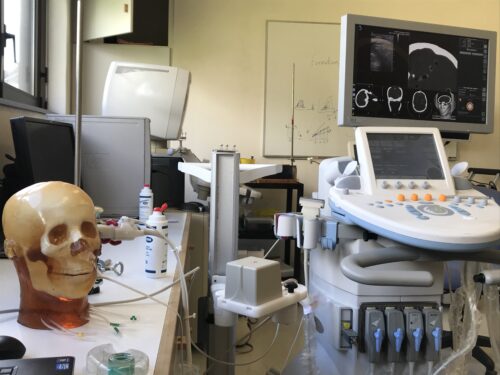
The research project Echobrain aims to innovate in transcranial ultrasound (US) imaging. Indeed, brain echography could be much more accessible than the current used techniques Xrays or Magnetic Resonance Imaging (MRI) but remains challenging due to strong attenuation and aberration of the US beam when crossing the skull bone. The quality of transcranial echography can be significantly increased provided that skull distortions can be accounted for at the emission, to focus the beam inside the brain, and at the reception, during the image formation process. These distortions can be experimentally measured, but need an invasive procedure generally using probes located inside the area to image. Models of US wave propagation, using a description of the morphology of the skull obtained by MRI or X-rays can also be used to predict these aberration corrections in a non-invasive way. From a scientific point of view, this project will provide a method to exhibit a simplified propagation model that suffices in order to obtain satisfactory correction of skull. From a technological point of view, it will contribute to the development of new US scanners dedicated to brain imaging based on programmable multichannel electronics. From a medical stand point, virtually all brain diseases could benefit from this improvement (stroke imaging at the early stage, cavitation activity monitoring, Doppler imaging, elastography of brain tumors or other lesions, intracranial pressure monitoring) The societal impact could therefore be significant.
